
Cholilithiyasis affects millions worldwide. The term cholilithiyasis refers to gallstone disease. It causes pain when stones block bile flow. Early detection reduces complications. This article defines cholilithiyasis, lists causes and risks, and explains symptoms, tests, and treatment options in 2026.
Table of Contents
ToggleKey Takeaways
- Cholilithiyasis, or gallstone disease, occurs when hardened deposits form in the gallbladder or bile ducts due to chemical imbalances in bile.
- Cholesterol and pigment stones are the main types of gallstones, forming from excess cholesterol or bilirubin respectively, often influenced by diet, genetics, and other risk factors.
- Early detection through ultrasound and other imaging tests is crucial for identifying gallstones and preventing complications like infection or bile duct obstruction.
- Risk factors for cholilithiyasis include high-fat diets, rapid weight loss, obesity, age, female sex, certain medications, and medical conditions affecting bile composition.
- Prevention strategies focus on a balanced diet, gradual weight loss, physical activity, and monitoring high-risk individuals with regular screening.
- Treatment ranges from observation for asymptomatic cases to laparoscopic cholecystectomy for recurrent symptoms, with urgent care needed for complicated cases presenting fever, jaundice, or severe pain.
What Is Cholelithiasis? Types Of Gallstones And How They Form
Cholilithiyasis describes the presence of gallstones in the gallbladder or bile ducts. Cholilithiyasis appears as hardened deposits that form from bile components. Cholilithiyasis forms when bile contains too much cholesterol, bilirubin, or when the gallbladder fails to empty properly.
There are two main types of gallstones in cholilithiyasis. Cholesterol stones appear as yellow-green stones that contain cholesterol crystals. Pigment stones appear as dark brown or black stones that contain excess bilirubin. Mixed stones contain both cholesterol and pigment components.
Cholilithiyasis begins with chemical imbalance in bile. Liver cells secrete bile that contains bile salts, cholesterol, and bilirubin. When cholesterol exceeds the solubility limit, crystals form. When bilirubin levels rise, pigment stones form. When the gallbladder fails to contract fully, bile sits and stones grow.
Small stones in cholilithiyasis may pass into the intestine without symptoms. Larger stones may lodge in the cystic duct or common bile duct. When a stone blocks a duct, bile and pancreatic enzymes back up. That blockage causes pain, inflammation, or infection. Chronic blockage can damage the gallbladder wall and surrounding organs.
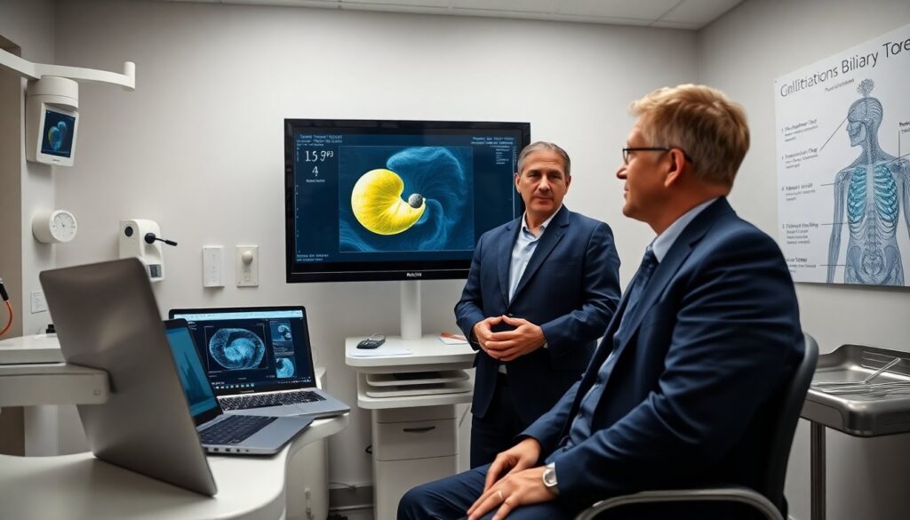
Modern imaging finds stones earlier in cholilithiyasis. Ultrasound shows most stones with high accuracy. CT and MRI provide extra detail when ultrasound is unclear. Blood tests do not detect stones directly but they point to infection or bile duct obstruction.
Causes, Risk Factors, And Prevention Strategies
Cholilithiyasis arises from a mix of causes. Genetics influence bile composition and gallbladder function. Diets high in saturated fat and low in fiber raise cholesterol levels and increase risk of cholilithiyasis. Rapid weight loss increases cholesterol saturation in bile and raises risk. Certain medical conditions, such as liver disease and hemolytic disorders, raise bilirubin and so risk of pigment stones in cholilithiyasis.
Age and sex affect cholilithiyasis risk. Older adults have higher risk. Women have higher risk due to estrogen effects on cholesterol metabolism. Pregnancy and hormonal therapy also raise the risk of cholilithiyasis. Obesity increases risk by raising cholesterol levels in bile.
Medications may increase risk of cholilithiyasis. Certain cholesterol-lowering drugs and hormone therapies can change bile composition. Total parenteral nutrition and prolonged fasting reduce gallbladder contractions and raise risk.
Prevention targets known risk factors for cholilithiyasis. A balanced diet with fiber and healthy fats lowers cholesterol levels. Gradual weight loss prevents rapid bile changes that favor stone formation. Regular physical activity helps maintain healthy weight and supports bile flow. When medications increase risk, clinicians review alternatives to lower cholilithiyasis chance.
Screening plays a role for high-risk groups. People with strong family history or chronic hemolysis may receive periodic ultrasound to monitor for cholilithiyasis. Vaccination and management of liver disease reduce secondary causes of pigment stones.
Symptoms, Diagnostic Tests, And When To Seek Medical Care
Symptoms of cholilithiyasis range from none to severe. Many people with cholilithiyasis remain asymptomatic. When symptoms appear, the most common sign is biliary colic. Biliary colic produces sudden, severe pain in the right upper abdomen or under the ribs. Pain often radiates to the back or right shoulder. Pain may last from minutes to several hours. Nausea and vomiting often accompany an attack.
Complicated cholilithiyasis causes fever, jaundice, or persistent severe pain. Fever with right upper quadrant pain suggests infection such as acute cholecystitis. Jaundice suggests obstruction of the common bile duct, which can lead to cholangitis or pancreatitis.
Clinicians diagnose cholilithiyasis with a focused approach. Ultrasound serves as the first-line test because it detects most stones and shows gallbladder inflammation. If ultrasound is inconclusive, magnetic resonance cholangiopancreatography (MRCP) or endoscopic ultrasound (EUS) provides clearer images of bile ducts. Blood tests measure white blood cell count, liver enzymes, bilirubin, and pancreatic enzymes to assess complications of cholilithiyasis.
When a bile duct stone is likely, endoscopic retrograde cholangiopancreatography (ERCP) both diagnoses and treats duct stones. ERCP removes stones with a side-viewing endoscope and instruments. Surgeons perform cholecystectomy to remove the gallbladder when cholilithiyasis causes recurrent symptoms. Laparoscopic cholecystectomy remains the standard surgical treatment in 2026.
Patients should seek urgent care for signs of complicated cholilithiyasis. They should seek care for fever plus abdominal pain, jaundice, severe ongoing pain, or vomiting that prevents oral intake. Early evaluation reduces the risk of severe infection, pancreatitis, and organ damage. Clinicians tailor treatment to the type of stone, severity of symptoms, and the patient’s overall health.